Metabolic regulation in pulmonary vascular disease
Pulmonary hypertension arising from chronic lung disease is common and carries the highest mortality of any subtype, yet only one therapy is approved for it and it helps only some patients. Sustained low oxygen thickens and narrows the pulmonary arteries, and that remodeling is driven by proliferation of the smooth muscle cells in the vessel wall. Most cells slow or stop dividing in hypoxia. Pulmonary artery smooth muscle cells do not, which implies a pathway that sustains growth when it should be suppressed.
The canonical account of hypoxic metabolism is that HIF-1α stabilizes, glycolytic gene expression rises, and glycolytic flux follows. Working in primary human lung fibroblasts and pulmonary artery smooth muscle cells rather than tumor lines, we found that this does not hold. Hypoxia decreased glycolysis in these cells even though HIF-1α was activated and glycolytic enzyme expression increased. Stabilizing HIF-1α pharmacologically in normal oxygen did raise glycolysis, but hypoxia blocked that response, so something downstream of HIF was overriding it. That something is MYC: knocking MYC down in hypoxia increased glycolysis, and overexpressing it in normoxia suppressed the glycolytic response to HIF stabilization. MYC signaling in hypoxia uncouples HIF-driven glycolytic transcription from glycolytic flux.
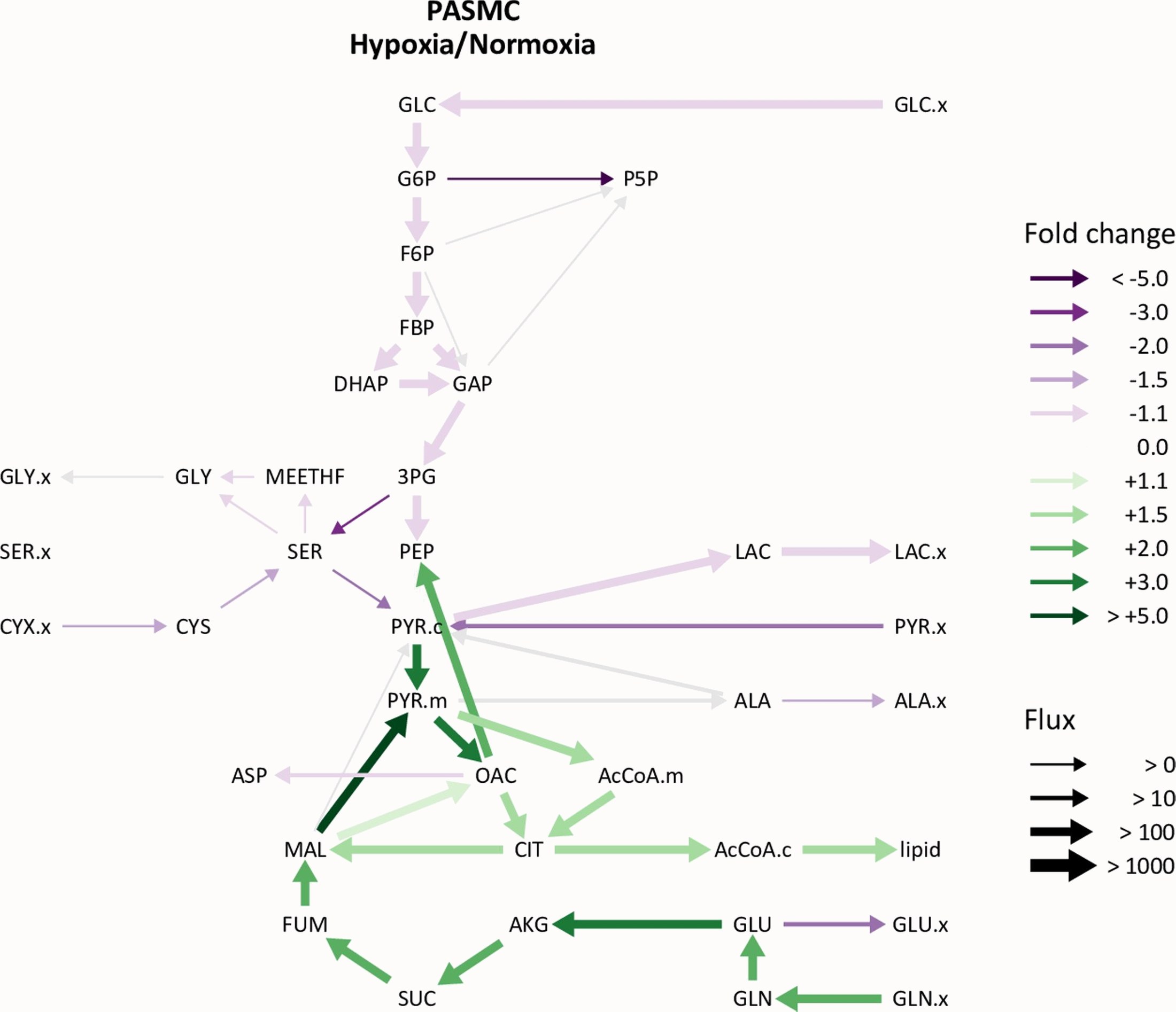
Modeled metabolic fluxes in pulmonary artery smooth muscle cells in low oxygen relative to room air.
Our current work asks what sustains MYC activity when oxygen is limited. In hypoxia, cells increasingly run the reaction catalyzed by isocitrate dehydrogenase in reverse, fixing carbon dioxide onto α-ketoglutarate to make citrate rather than oxidizing citrate to α-ketoglutarate. This reductive carboxylation supplies carbon for biosynthesis when oxidative metabolism is constrained, and our preliminary data indicate that it is required to maintain MYC signaling and proliferation in hypoxic smooth muscle cells.
This gives a metabolic–transcriptional axis to test. We are quantifying IDH expression in remodeled human pulmonary arteries and asking whether inhibiting IDH — with compounds already approved in oncology — suppresses reductive carboxylation, MYC activation, and proliferation. In parallel, inducible smooth-muscle-specific expression of Omomyc, a peptide inhibitor of MYC now in clinical trials, will test whether MYC is required for hypoxic pulmonary vascular remodeling in an intact animal. Because both classes of drug are already well advanced in human medicine, a positive result would have a short path to trial in a patient population with few options.
Key paper
Data and code
- Raw data and figure code — research compendium, archived at Software Heritage
- RNA-seq reads — NIH SRA, PRJNA721596
- Prolyl hydroxylase inhibition RNA-seq — molidustat-treated lung fibroblasts